In the previous part, we had a look at the brain size and growth rate of autistic children, which was found to be increased at a young age; in fact, at age 3–5, the autistic brain weighs the same as the brain of the average neurotypical adult male.
Now we will have a look at the brain differences in the cerebrum. I will explain what the cerebrum is and does first, so if you already know this, feel free to skip the next part.
What is the cerebrum?
The brain has three main parts: the brainstem, the cerebellum, and the cerebrum.
These brain parts are layered like an onion, with the cerebrum being the outermost part, composed of left and right cerebral hemispheres. The cerebrum gives us higher cognition, and is involved in thinking, perceiving, understanding, and language.
The cerebrum is basically the processor of the brain; it processes and integrates sensory information and neural functions to give us senses, thoughts, and movements.
 Cerebral lobes
The cerebrum is divided into four regions called lobes:
- The frontal lobe, which is associated with reasoning, planning, parts of speech, movement, emotions, and problem-solving.
- The temporal lobe, which is associated with perception and recognition of auditory stimuli, memory, and speech.
- The parietal lobe, which is associated with movement, orientation, recognition, and perception of stimuli.
- The occipital lobe, which is associated with visual processing.
In upcoming posts in this series, we will go into more detail on these subregions, and the differences found in the autistic brain.
But now that you (approximately) understand what the cerebrum is and does, let’s have a look at what is different about the autistic information processor.
Cerebral volume
Finding: Increases in cerebral volume[1]Magnetic Resonance Imaging and Head Circumference Study of Brain Size in Autism[2]Unusual brain growth patterns in early life in patients with autistic disorder: an MRI study[3]Brain structural abnormalities in young children with autism spectrum disorder
Method: Magnetic resonance imaging (MRI)
Based on the findings of increased head circumference in autistic children[4]Head circumference and height in autism: A study by the collaborative program of excellence in autism researchers made inferences about the timing of brain enlargements.
A study from 2005 found generalized enlargement of white matter and gray matter volumes in the cerebrum in autistic children at 2 years of age. These increases in white and gray matter were not found in the cerebellum.*[5]Magnetic Resonance Imaging and Head Circumference Study of Brain Size in Autism
- Actually, an earlier study from 2002 already indicated that the volume of the cerebrum is significantly increased in autistic children, and they did find volume increases in the cerebellum as well, but no disproportional increase; in other words, our cerebellum is larger in size, but proportional to the cerebral volume as a whole.[6]Brain structural abnormalities in young children with autism spectrum disorder
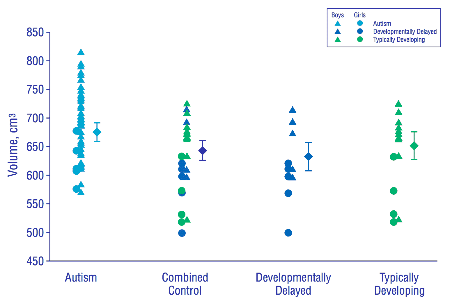
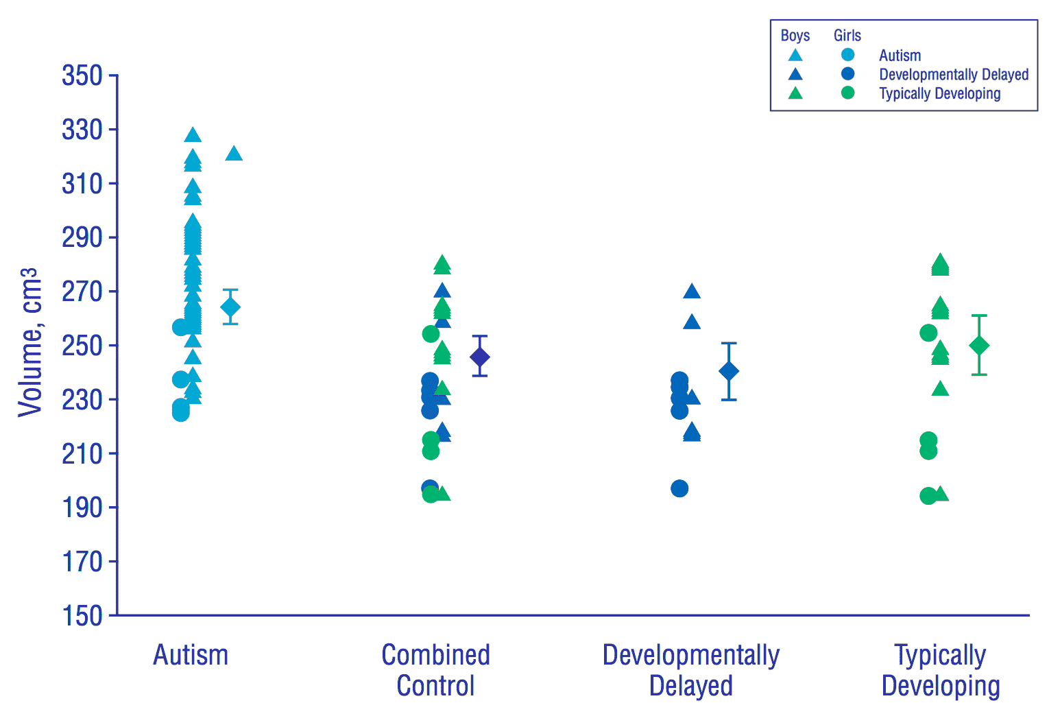
In the diagrams below you can see how gray and white matter volumes are significantly increased in autistics compared with typically developing children, and in particular developmentally delayed children. So while some autistic children may experience certain delays in development, at least in terms of gray and white matter volumes in the cerebrum, autistic children are clearly distinct from developmentally delayed children.
Gray matter volume in the cerebrum

White matter volume in the cerebrum

Indirect evidence suggests that this increased rate of brain growth in autism occurs in the latter part of the 1st year of life. This is also in accordance with the research we talked about in the previous post.
Language disorder
Research from 2004 indicates that white matter volume enlargements are seen in both autism and developmental language disorder (DLD), which suggests the two conditions are somehow related.[7]Localization of white matter volume increase in autism and developmental language disorder
In the Venn diagram below, you can see DLD and related disorders that are part of speech, language, and communication needs (SLCN).
Speech, language, and communication needs
- For more information on the statement ‘language disorder associated with biomedical condition X’ in the infographic above, read Statement 6 of the consensus paper Phase 2 of CATALISE: a multinational and multidisciplinary Delphi consensus study of problems with language development: Terminology.
In the Venn diagram below you can see autism is actually considered a language disorder by some criteria, although not necessarily part of the SLCN construct.
Speech, language, and communication needs (incl. ASD)
Note however that delays in communication and language development are not necessarily inherent to autism; not every autistic person has experienced delays in these areas. So what an increase of white matter means for the entire autism spectrum, and to what extent it is linked to DLD, I do not know.
Hemispheric connections
The study from 2004 also indicated autism and DLD are linked in the effect white matter has on the brain:
These findings suggest an ongoing postnatal process in both autism and DLD that is probably intrinsic to white matter, that primarily affects intra-hemispheric and corticocortical connections, and that places these two disorders on the same spectrum.[8]Localization of white matter volume increase in autism and developmental language disorder
What the quote above means is that both in autism and DLD a process occurs that is likely linked to the white matter increases during development after birth, which affects the connections within hemispheres, as well as the connections between cortexes. In other words, this alludes to the localized hyperconnectivity that we talked about in the first part of this series about connectivity:
Autistic brain differences pt. 1 – Connectivity
Gray matter
Finding: Gray matter increases in regions related to social cognition, communication, repetitive behaviors, and auditory & visual perception[9]Neuroanatomical differences in brain areas implicated in perceptual and other core features of autism revealed by cortical thickness analysis and voxel-based morphometry
Method: MRI
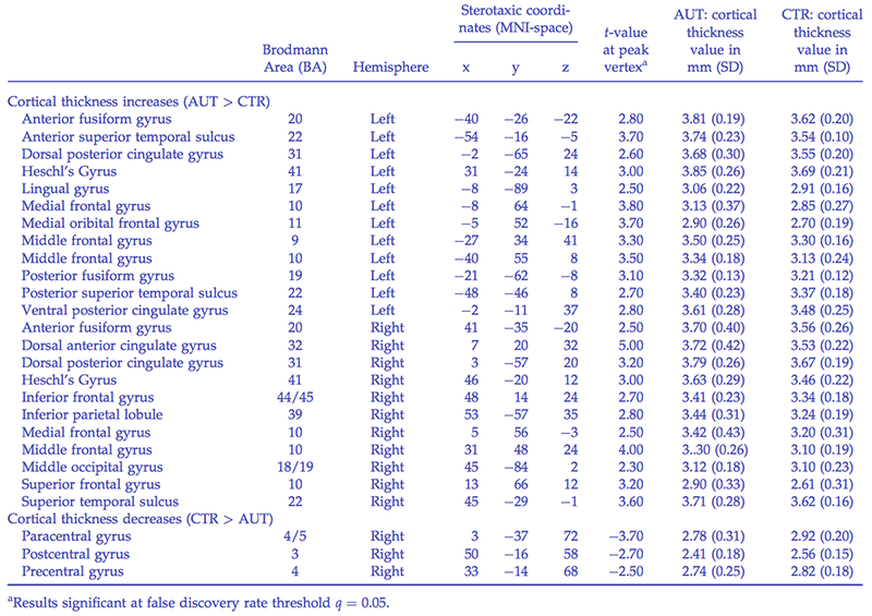
Analysis of the thickness of the cortexes, (the outermost layer of an organ, in this case of the brain) and voxel-based morphometry techniques both revealed structural brain differences (mostly in terms of gray matter increases) in brain areas implicated in social cognition, communication, and repetitive behaviors (the three core domains of autism).[10]Neuroanatomical differences in brain areas implicated in perceptual and other core features of autism revealed by cortical thickness analysis and voxel-based morphometry
Group cortical thickness differences

Cerebral cortex
So the cortical thickness of certain brain regions is linked to the three core domains of autism. I think the implication of the research is thus that there is a link to the challenges autistic people tend to have in these core domains, but it’s important to note that these brain differences are not solely linked to challenges, but autistic differences in general—including certain aptitudes.
For instance, the cerebral cortex—the outermost layer of the cerebrum—plays a key role in memory, attention, perceptual awareness, thought, language, and consciousness. Based on personal observations as well as a wealth of research—including the very table above which details the many regions of the cerebral cortex that are thicker than in neurotypicals—it is far from uncommon for autistic people to excel in one or several of these areas. Read for example our post below on savant syndrome:
Autism & savant syndrome
Primary auditory cortex
The researchers of the 2004 study mention the cortical thickness increase of the primary auditory cortex in the autistic brain as a particular area of interest given the evidence on enhanced low-level pitch perception of autistic individuals.
The cortical thickness increase found in the primary auditory cortex of the autistic group is particularly interesting given that this region is known to play a key role in low-level pitch perception in typically developing individuals (McDermott and Oxenham, 2008; Zatorre, 2001), and individuals with autism have enhanced low-level pitch perception (Bonnel et al., 2003;[11]Enhanced Pitch Sensitivity in Individuals with Autism: A Signal Detection Analysis Heaton, 2005;[12]Interval and Contour Processing in Autism Heaton et al., 2008;[13]Superior discrimination of speech pitch and its relationship to verbal ability in autism spectrum disorders Samson et al., 2006[14]Emotional responses to unpleasant music correlates with damage to the parahippocampal cortex).[15]Neuroanatomical differences in brain areas implicated in perceptual and other core features of autism revealed by cortical thickness analysis and voxel-based morphometry
So as you can see, while the differences found in the cerebrum play an essential role in some of our challenges, it is equally linked to some amazing abilities, some of which are listed on our Powers & Kryptonite page.
Comments
Let us know what you think!